Home

kıvrılmış hastalık dağ cholesteatoma diffusion mri kıyıya yakın inci gibi kapanıyorum

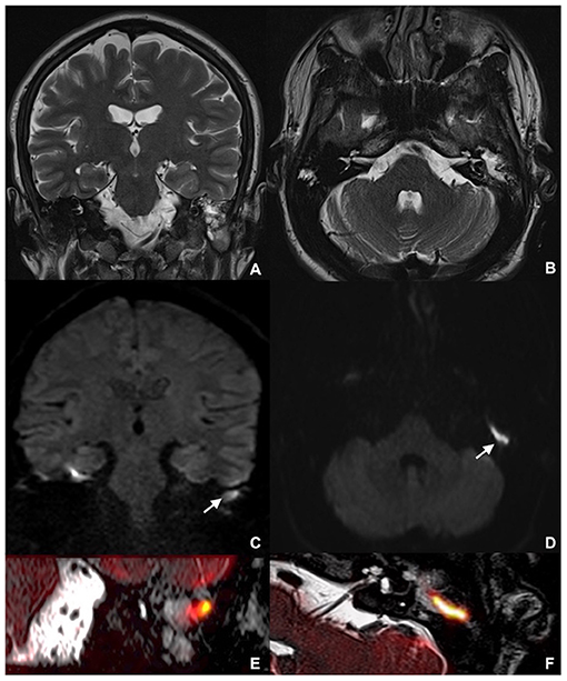
Frontiers | Combining Thin-Section Coronal and Axial Diffusion Weighted  Imaging: Good Practice in Middle Ear Cholesteatoma Neuroimaging
Frontiers | Combining Thin-Section Coronal and Axial Diffusion Weighted Imaging: Good Practice in Middle Ear Cholesteatoma Neuroimaging

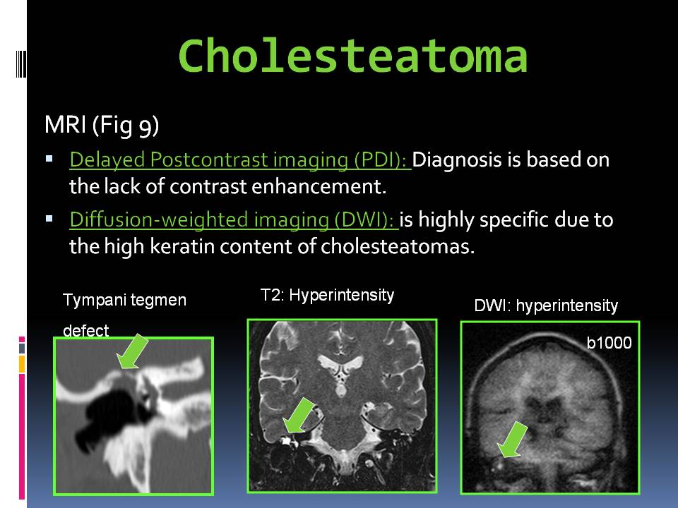
Dr Balaji Anvekar FRCR: Role of Diffusion imaging in Cholesteatoma  evaluation
Dr Balaji Anvekar FRCR: Role of Diffusion imaging in Cholesteatoma evaluation

Diffusion-Weighted MR Imaging of Cholesteatoma in Pediatric and Adult  Patients Who Have Undergone Middle Ear Surgery | AJR
Diffusion-Weighted MR Imaging of Cholesteatoma in Pediatric and Adult Patients Who Have Undergone Middle Ear Surgery | AJR

The Clinical Role of Diffusion-Weighted MRI for Detecting Residual  Cholesteatoma in Canal Wall up Mastoidectomy | Indian Journal of  Otolaryngology and Head & Neck Surgery
The Clinical Role of Diffusion-Weighted MRI for Detecting Residual Cholesteatoma in Canal Wall up Mastoidectomy | Indian Journal of Otolaryngology and Head & Neck Surgery

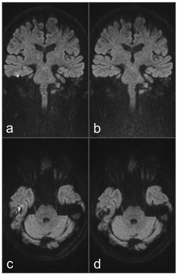
Detection of Middle Ear Cholesteatoma by Diffusion-Weighted MR Imaging:  Multishot Echo-Planar Imaging Compared with Single-Shot Echo-Planar Imaging  | American Journal of Neuroradiology
Detection of Middle Ear Cholesteatoma by Diffusion-Weighted MR Imaging: Multishot Echo-Planar Imaging Compared with Single-Shot Echo-Planar Imaging | American Journal of Neuroradiology

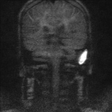
Cholesteatoma(DWI IAM'S) MRI Protocols and Planning | Indications for  Diffusion Weighted IAM's Scan
Cholesteatoma(DWI IAM'S) MRI Protocols and Planning | Indications for Diffusion Weighted IAM's Scan

Rapid diffusion-weighted MRI for the investigation of recurrent temporal  bone cholesteatoma - Richard G Kavanagh, Stephen Liddy, Anne G Carroll,  Yvonne M Purcell, Anna E Smyth, S Guan Khoo, Graeme McNeill, Dermot
Rapid diffusion-weighted MRI for the investigation of recurrent temporal bone cholesteatoma - Richard G Kavanagh, Stephen Liddy, Anne G Carroll, Yvonne M Purcell, Anna E Smyth, S Guan Khoo, Graeme McNeill, Dermot

Diffusion Weighted MR Imaging of Primary and Recurrent Middle Ear  Cholesteatoma: An Assessment by Readers with Different Expertise
Diffusion Weighted MR Imaging of Primary and Recurrent Middle Ear Cholesteatoma: An Assessment by Readers with Different Expertise

Recurrent cholesteatoma. A , Echo-planar DWI shows artifacts ( double... |  Download Scientific Diagram
Recurrent cholesteatoma. A , Echo-planar DWI shows artifacts ( double... | Download Scientific Diagram

JPM | Free Full-Text | The Efficacy of DW and T1-W MRI Combined with CT in  the Preoperative Evaluation of Cholesteatoma
JPM | Free Full-Text | The Efficacy of DW and T1-W MRI Combined with CT in the Preoperative Evaluation of Cholesteatoma

Diffusion-Weighted Magnetic Resonance Imaging of Cholesteatoma Using  PROPELLER at 1.5T: A Single-Centre Retrospective Study - ScienceDirect
Diffusion-Weighted Magnetic Resonance Imaging of Cholesteatoma Using PROPELLER at 1.5T: A Single-Centre Retrospective Study - ScienceDirect

The Utility of Diffusion-Weighted Imaging for Cholesteatoma Evaluation |  American Journal of Neuroradiology
The Utility of Diffusion-Weighted Imaging for Cholesteatoma Evaluation | American Journal of Neuroradiology
EPOS™
EPOS™

Diagnostics | Free Full-Text | Comparison of Diagnostic Performance and  Image Quality between Topup-Corrected and Standard Readout-Segmented  Echo-Planar Diffusion-Weighted Imaging for Cholesteatoma Diagnostics
Diagnostics | Free Full-Text | Comparison of Diagnostic Performance and Image Quality between Topup-Corrected and Standard Readout-Segmented Echo-Planar Diffusion-Weighted Imaging for Cholesteatoma Diagnostics

Non-echoplanar diffusion weighted imaging in the detection of  post-operative middle ear cholesteatoma: navigating beyond the pitfalls to  find the pearl | Insights into Imaging | Full Text
Non-echoplanar diffusion weighted imaging in the detection of post-operative middle ear cholesteatoma: navigating beyond the pitfalls to find the pearl | Insights into Imaging | Full Text

Cholesteatoma(DWI IAM'S) MRI Protocols and Planning | Indications for  Diffusion Weighted IAM's Scan
Cholesteatoma(DWI IAM'S) MRI Protocols and Planning | Indications for Diffusion Weighted IAM's Scan

Surgically confirmed cholesteatoma. a Non-EP DWI showing a high... |  Download Scientific Diagram
Surgically confirmed cholesteatoma. a Non-EP DWI showing a high... | Download Scientific Diagram

Cholesteatoma | Radiology Case | Radiopaedia.org

Performance of TGSE BLADE DWI compared with RESOLVE DWI in the diagnosis of  cholesteatoma | BMC Medical Imaging | Full Text
Performance of TGSE BLADE DWI compared with RESOLVE DWI in the diagnosis of cholesteatoma | BMC Medical Imaging | Full Text
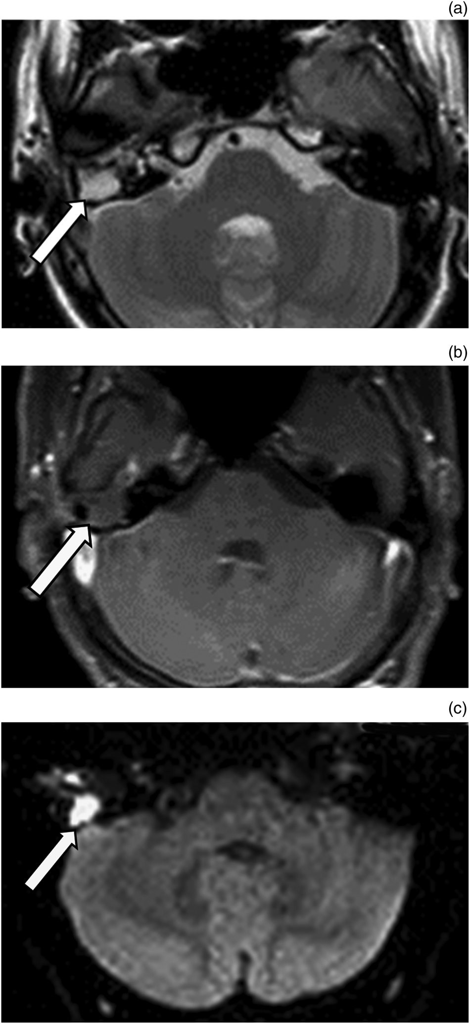
Reliability of diffusion-weighted magnetic resonance imaging in  differentiation of recurrent cholesteatoma and granulation tissue after  intact canal wall mastoidectomy | The Journal of Laryngology & Otology |  Cambridge Core
Reliability of diffusion-weighted magnetic resonance imaging in differentiation of recurrent cholesteatoma and granulation tissue after intact canal wall mastoidectomy | The Journal of Laryngology & Otology | Cambridge Core

Figure 1 from PROPELLER non-EPI DWI in the diagnosis of primary and  recurrent cholesteatoma . A pictorial review | Semantic Scholar
Figure 1 from PROPELLER non-EPI DWI in the diagnosis of primary and recurrent cholesteatoma . A pictorial review | Semantic Scholar

Contemporary Non–Echo-planar Diffusion-weighted Imaging of Middle Ear  Cholesteatomas | RadioGraphics
Contemporary Non–Echo-planar Diffusion-weighted Imaging of Middle Ear Cholesteatomas | RadioGraphics

The role of magnetic resonance imaging in the postoperative management of  cholesteatomas | Brazilian Journal of Otorhinolaryngology
The role of magnetic resonance imaging in the postoperative management of cholesteatomas | Brazilian Journal of Otorhinolaryngology

A 41-year-old male with a large cholesteatoma on the left side. MR... |  Download Scientific Diagram
A 41-year-old male with a large cholesteatoma on the left side. MR... | Download Scientific Diagram

BIR Publications
BIR Publications

The Utility of Diffusion-Weighted Imaging for Cholesteatoma Evaluation |  American Journal of Neuroradiology
The Utility of Diffusion-Weighted Imaging for Cholesteatoma Evaluation | American Journal of Neuroradiology

The Utility of Diffusion-Weighted Imaging for Cholesteatoma Evaluation |  American Journal of Neuroradiology
The Utility of Diffusion-Weighted Imaging for Cholesteatoma Evaluation | American Journal of Neuroradiology